Treatment of Retinal Tumors by Ophthalmic Artery Catheterization
Treatment of Retinal Tumors by Ophthalmic Artery Catheterization
Naturally, the iris is black and may appear red during photography, which is normal. However, a white iris, or the regular red reflex, is not a normal condition.
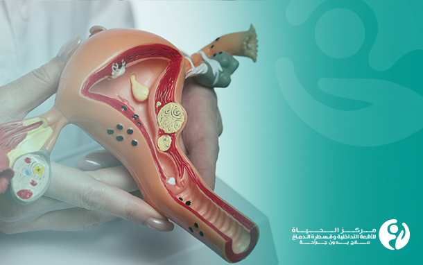
Retinoblastoma is a rare tumor of the eye that typically occurs in children and is a malignant (cancerous) tumor that affects the retina. Retinoblastoma arises from a mutation in the gene that controls how cells divide, so cells in the retina grow uncontrollably and become cancerous.
Retinoblastoma is a complex disease that requires precise diagnostic work and a multidisciplinary expert team for appropriate treatment, improving survival while preserving vision and long-term monitoring; this is achieved at Al Hayat Center for Interventional Radiology and Neurointervention in Iraq, where chemotherapy is injected directly into the ophthalmic artery using catheterization instead of enucleation (removal of the eye) or systemic intravenous chemotherapy, which helps achieve better benefit and fewer complications.
Facts about retinoblastoma:
• Retinoblastoma is the most common primary intraocular malignant tumor in children.
• Retinoblastoma represents 3% of cancer cases in children under the age of 15.
• The tumor size can range from very small to large enough to destroy the entire eye.
• Retinoblastoma may be unilateral (in one eye) or bilateral (in both eyes).
• The tumor may be located within the eyeball or extend along the optic nerve. In rare cases, retinoblastoma may spread within the skull.
• Retinoblastoma can be hereditary or non-hereditary. The hereditary type is usually diagnosed before the age of 1 year, while the non-hereditary type is diagnosed by the age of 2 years.
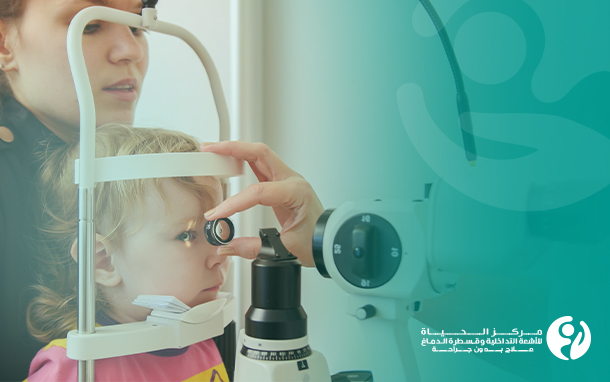
• Typically, retinoblastoma is first noticed by a parent or pediatrician who sees a white iris in a child's eye.
• Prompt diagnosis is crucial for saving life, the eye, and vision.
• Interventional radiologists play an important role in diagnosing, staging, and treating retinoblastoma without eye removal.
Symptoms of retinoblastoma (Eye Tumor):
• Strabismus.
• Double vision.
• Pain and redness in the eye.
• Decreased vision.
• Difference in iris color between the two eyes.
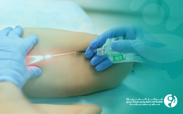
The ideal treatment for children to save the affected eye is intra-arterial chemotherapy in the interventional radiology rooms.
Intra-arterial chemotherapy injection for retinoblastoma:
Technological advancements and modern techniques allow selective chemotherapy injection through the ophthalmic artery (which supplies blood to the eye), achieving a healing rate of over 70% for advanced retinoblastoma with minimal systemic side effects and toxicity.
History of retinoblastoma treatment:
The treatment of intraocular retinoblastoma has improved significantly over the past century. It is the most common intraocular tumor in childhood, which was previously considered a fatal cancer for children. However, modern treatment approaches have achieved success stories, with a survival rate of more than 99%. In the past, the goal of treating advanced intraocular retinoblastoma was to save the child's life, while current therapies have made it possible to target eye preservation and visual acuity. One of the most remarkable modern treatment approaches is super-selective intra-arterial chemotherapy, also known as ophthalmic artery chemosurgery (OAC).
The evolution of retinoblastoma treatment to ophthalmic artery chemotherapy:
In 1954, intra-arterial chemotherapy was first performed to treat retinoblastoma by using chemotherapy to reduce the total dose of external beam radiation therapy required to treat retinoblastoma.
Since 1988, a type of intra-arterial chemotherapy has been used to treat retinoblastoma. However, this method is not selective, as there are nearby branches where the drug can flow before reaching the ophthalmic artery, and it was aimed at providing a treatment option for families who wanted to avoid eye removal at all costs, not to reduce the dose.
In 2006, a microcatheter was precisely placed through the femoral artery under direct fluoroscopic visualization into the ophthalmic artery, allowing selective delivery of chemotherapy to the eye. Following the surgery, patients are observed for four to six hours. Currently, more than 30 countries use this strategy.
One of the motivations for improving the intra-arterial injection technique was to deliver a higher concentration of chemotherapy without increasing side effects in the hope of successfully treating advanced disease. Studies have shown that OAC can be used successfully as a first-line treatment. It has been used to treat less advanced diseases, advanced diseases in young infants, and cases of retinal detachment. Studies have also shown its effectiveness in saving eyes, as the intra-arterial chemotherapy delivers a very high dose directly to the tumor, increasing the treatment efficacy and minimizing side effects to the minimum. Therefore, Warith International Cancer Institute sought to implement it for the first time in Iraq at the Al Hayat Center for Interventional Radiology and Neurointervention in Karbala, with the highest success rates by qualified doctors to serve Iraqi children.
Why choose the Al Hayat Center for Interventional Radiology and Neurointervention?
• Accurate diagnosis is guaranteed with the latest equipment available at the Al Hayat Center for Interventional Radiology and Neurointervention. The importance of the diagnostic steps lies in the need for detailed imaging to verify the presence of a tumor inside the skull. It is also necessary to carefully evaluate the other eye through imaging, especially in children with hereditary retinoblastoma. Evaluation of the other eye requires great precision because any tumor on the opposite side is usually much smaller than the tumor of the symptomatic eye.
• An integrated medical team is required before, during, and after the procedure. The close coordination between the ocular oncologist and the interventional radiologist is the first step to ensure success.
• The vast majority of these patients are under 3 years of age, so their blood vessels are very small and fragile. Therefore, it is essential to rely on an expert interventional radiologist who can perform the procedures safely for these children, which Al Hayat Center for Interventional Radiology and Neurointervention in Iraq provides.
• Thorough preparation is essential. For example, many interventional radiologists may be able to access the child's blood vessels, but to what extent should the catheter be inserted into the ophthalmic artery, and how exactly should the chemotherapy be infused? Therefore, a detailed plan must be developed before offering this treatment as a therapeutic option.
Retinoblastoma is the most common primary intraocular tumor in childhood. Accurate early diagnosis is important to achieve maximum patient survival and save the eye and visual function. A multidisciplinary team is required to improve patient outcomes. The interventional radiologist is a key member of the retinoblastoma care team, with intra-arterial chemotherapy as an ideal care option to save the eye; thus, Al Hayat Center for Interventional Radiology and Neurointervention plays an active role in the management of retinoblastoma.